-
所属(所属キャンパス)
-
医学部 共同利用研究室(細胞組織学研究室) ( 信濃町 )
-
職名
-
教授(有期)
-
外部リンク
-
-
松尾 光一 ( マツオ コウイチ )
Matsuo, Koichi
|
|
|
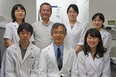
細胞組織学研究室のメンバー(2017)
研究室は、破骨細胞、さらに破骨細胞と骨芽細胞の相互作用(カップリング)に関する一連の骨代謝学の研究で成果を挙げた。近年では、骨の三次元的な形態形成に研究の焦点を移し、内軟骨性骨化や生後の骨形態変化のメカニズムを、マウスやニホンザルなどの実験動物を用いて解析している。「多様なサイズとかたち」を生み出し、それを成長させ、維持し修復する細胞レベルのメカニズムは本質的なものであるにもかかわらず、不明な点が多い。新たな研究領域を開拓しようと、長管骨や椎骨に加え、耳小骨や頭蓋底骨などを対象として解析力を高めている。さらに放射光施設SPring-8(兵庫)におけるX線位相顕微鏡によるCT撮影(東北大学との共同研究)では、 細胞レベルの解像度で、内軟骨性骨化を担う骨形成性毛細血管の構造を明らかにした。研究室を挙げて形態形成原理の追究に精力を注いでいる。
国立長寿医療研究センター老年病研究部, 室長
慶應義塾大学医学部, 微生物学・免疫学教室, 助教授
慶應義塾大学, 微生物学・免疫学教室, 准教授
慶應義塾大学医学部, 共同利用研究室(細胞組織学), 教授
総合医科学研究センター長
骨代謝学
骨免疫学
ライフサイエンス / 細胞生物学 (Cell Biology)
ライフサイエンス / 解剖学 (General Anatomy(includes Histology/Embryology))
内軟骨性骨化における骨形成性血管の解析,
骨の形態形成とバイオミネラリゼーション・恒常性維持のメカニズムを、細胞間相互作用によって解明することを目指している。,
Trans-pairing between osteoclasts and osteoblasts shapes the cranial base during development
Edamoto M., Kuroda Y., Yoda M., Kawaai K., Matsuo K.
Scientific Reports (Scientific Reports) 9 ( 1 ) 2019年12月
Yang M., Arai A., Udagawa N., Zhao L., Nishida D., Murakami K., Hiraga T., Takao-Kawabata R., Matsuo K., Komori T., Kobayashi Y., Takahashi N., Isogai Y., Ishizuya T., Yamaguchi A., Mizoguchi T.
Journal of Bone and Mineral Research (Journal of Bone and Mineral Research) 34 ( 10 ) 1952 - 1963 2019年10月
ISSN 08840431
Innervation of the tibial epiphysis through the intercondylar foramen
Matsuo K., Ji S., Miya A., Yoda M., Hamada Y., Tanaka T., Takao-Kawabata R., Kawaai K., Kuroda Y., Shibata S.
Bone (Bone) 120 297 - 304 2019年03月
ISSN 87563282
Bone Marrow Cells Inhibit BMP-2-Induced Osteoblast Activity in the Marrow Environment
Nguyen H.T., Ono M., Oida Y., Hara E.S., Komori T., Akiyama K., Nguyen H.T.T., Aung K.T., Pham H.T., Tosa I., Takarada T., Matsuo K., Mizoguchi T., Oohashi T., Kuboki T.
Journal of Bone and Mineral Research (Journal of Bone and Mineral Research) 34 ( 2 ) 327 - 332 2019年02月
ISSN 08840431
Sasaki M., Chubachi S., Kameyama N., Sato M., Haraguchi M., Miyazaki M., Takahashi S., Nakano T., Kuroda Y., Betsuyaku T., Matsuo K.
PLoS ONE (PLoS ONE) 13 ( 1 ) 2018年01月
ISSN 1932-6203
骨細胞ネットワークによるバイオミネラリゼーション
松尾光一
Clinical calcium 25 ( 10 ) 1461 - 1466 2015年10月
記事・総説・解説・論説等(商業誌、新聞、ウェブメディア), 単著, ISSN 0917-5857
骨細胞による骨融解に対するグルココルチコイドの影響
松尾 光一
Clinical calcium 24 ( 9 ) 1337 - 1342 2014年09月
記事・総説・解説・論説等(商業誌、新聞、ウェブメディア), 単著, ISSN 0917-5857
骨免疫から見た老化制御
松尾光一
Clinical calcium 23 ( 1 ) 59 - 64 2013年01月
記事・総説・解説・論説等(商業誌、新聞、ウェブメディア), 単著, ISSN 0917-5857
Eph-ehrinファミリー分子群による骨代謝制御
Matsuo K、, Otaki N
Clinical calcium 22 ( 11 ) 1669 - 1675 2012年11月
記事・総説・解説・論説等(商業誌、新聞、ウェブメディア), 共著, ISSN 0917-5857
骨細胞による骨融解:骨小腔体積の計測
松尾光一
Clinical calcium 22 ( 5 ) 677 - 683 2012年05月
記事・総説・解説・論説等(商業誌、新聞、ウェブメディア), ISSN 0917-5857
Mammalian-type Brn-1/Pou3f3 with 'homopolymeric amino acids (HPAAs)' is essential for stress-induced bone metabolic changes in mice
松尾 光一
[国際会議] Australian New Zealand Bone & Mineral Society Annual Scientific Meeting (Hobart, Tasmania) ,
ポスター発表
Seasonality in Bone Mineralization of Auditory Ossicles and Long Bones in the Primate Macaca fuscata
松尾 光一
[国際会議] Annual Meeting of the American Society for Bone and Mineral Research (Seattle, Washington, USA) ,
ポスター発表
Sialylated Glycans of MMP-9 Marak Bone Resorption Lacunae.
松尾 光一
[国際会議] Annual Meeting of the American Society for Bone and Mineral Research (Seattle, Washington, USA) ,
ポスター発表
Continuous Parathyroid Hormone Injection in Mouse Has Differential Effects on Osteoclast Activation in Primary and Secondary Spongiosa.
松尾 光一
[国際会議] Annual Meeting of the American Society for Bone and Mineral Research (Seattle, Washington, USA) ,
ポスター発表
EphB/ephrin-B interactions regulate stromal cell fate determination and bone marrow support
松尾 光一
[国際会議] Annual Meeting of the American Society for Bone and Mineral Research (Seattle, Washington, USA) ,
ポスター発表
ホモキラル要素による左右相称骨格の構築技法
文部科学省・日本学術振興会, 科学研究費助成事業, 松尾 光一, 学術変革領域研究(A), 補助金, 研究代表者
左右対称性の骨格が形成される細胞機構の理解に向けて
文部科学省・日本学術振興会, 科学研究費助成事業, 松尾 光一, 基盤研究(B), 補助金, 研究代表者
骨形成性毛細血管からみた内軟骨性骨化の新しい概念
文部科学省・日本学術振興会, 科学研究費助成事業, 松尾 光一, 基盤研究(B), 補助金, 研究代表者
日本骨代謝学会学術賞
2014年07月, 日本骨代謝学会
北里賞
2003年06月, 慶應義塾大学医学部
Young Investigator Award
1997年09月, American Society for Bone and Mineral Research
分子細胞生物学
2025年度
微生物学
2025年度
メディカル・プロフェッショナリズムⅢ
2025年度
医学概論
2025年度
学外特別研修(インターンシップ)
2025年度
微生物学 (細菌学各論 1,2)
慶應義塾
春学期, 講義
メディカル・プロフェッショナリズム III (研究倫理)
慶應義塾
春学期, 講義
基礎分子細胞生物学 II
慶應義塾
春学期, 講義
微生物学 (細菌学各論 1,2)
慶應義塾
春学期, 講義
メディカル・プロフェッショナリズム III (研究倫理)
慶應義塾
春学期, 講義


Click to view the Scopus page. The data was downloaded from Scopus API inFebruary 09, 2026, via http://api.elsevier.com and http://www.scopus.com .